Projects |
||
|
|
|
[ Home ] [ Projects ] [ Links ] [ Biography ] [ Publications ]
Interested in a BSc or MSc end project? We always have student projects available. Contact me by email or simply walk into my office (F256). |
If you are looking for a PhD or PostDoc position, write an email with your CV and a motivation letter. We are always looking for enthousiastic young people wanting to advance science and technology! |
||||||||||||||||||||
Precise to the nanometerThe position of stochastically blinking fluorescent molecules can be measured with nanometer precision from a movie acquired with a conventional widefield microscope. A super-resolution image can be reconstructed from the localization data, thereby circumventing the classical diffraction barrier to resolution. We develop new imaging modalities in this area (measuring emission colour and molecular orientation along with emitter position) and work on image analysis methods for image quantitation, for establishing performance limits, and for optimizing image acquisition and reconstruction protocols. |
Shining lightAn intriguing way to make a 3D fluorescence image is to record a set of (2D) images on a camera for a set of specifically designed illumination patterns. As a bonus the in-plane resolution can be doubled as well. The design of illumination patterns in combination with various ways of scanning these patterns and image analysis methods can improve light efficiency and robustness.
|
||||||||||||||||||||
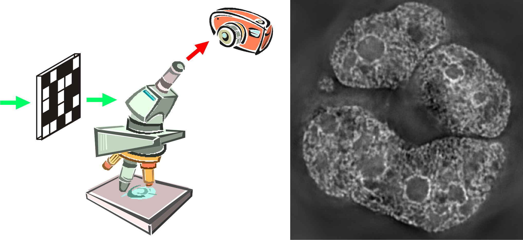
Pathology going digitalDigital pathology is an emerging clinical practice in which a pathologist makes a diagnosis by examining a digital high-resolution image of a tissue slide. These images are acquired with a high-throughput automated microscope ("whole slide scanner"). We develop efficient optical quality testing methods for inspection of manufacturing quality and for monitoring systems during their operational lifetime, we work on new ways for scanning multiple focal slices simultaneously, and we investigate image analysis algorithms for diagnostic assistance.
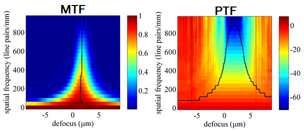
The top image shows an example of an image taken with our whole slide scanning setup, the bottom images show a measurement of the through-focus Modulation Transfer Function and Phase Transfer Function. |
|||||||||||||||||||||
PhD Theses
|
|||||||||||||||||||||