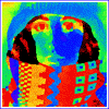
 Two chromosome probes + nucleus counterstain
The use of FISH technology (fluorescence in situ hybridization) requires the analysis of a large number of interphase cells. The analysis consists of finding the cell nuclei and counting the number of hybridization spots in the nuclei. In a clinical setting it is necessary to review 500 to 1000 cells in order to determine the distribution of spots per cell and to be able to detect small aberrant sub-populations of cells. Practical considerations requires that such a test can be performed in a short time (< 20 minutes).
We have developed a completely automated system that counts hybridization spots for one probe in an interphase cell nuclei [1,2]. The system is built around a Leitz Aristoplan microscope, equipped with motorized stage, focus control, and a Photometrics KAF 1400 camera. A Macintosh 840 AV controls the screening and analyzes the images. Slides are screened with 25x/0.75 NA objective and a dual bandpass filter block. A complete scanning cycle consists of automated focusing of the field, acquisition of the image, processing the image, and then moving the stage to the next field. This continues until a preset number of cells has been counted.
To analyze 500 cells, 100 to 500 Mbytes of data - depending on the cell density on the slide - have to be acquired and processed. In order to achieve this in a reasonable amount of time, each step must be optimized with respect to speed. The auto-focusing algorithm [3] is applied on 2x4 binned image and uses extrapolation to stay in focus without examining each field. The image processing starts with sub-sampling the image by a factor of 8 in both directions. This sub-sampled image is used to find regions of interest that contain single nuclei. Because only a small percentage of the area contains cells, this first step significantly reduces the amount of data and is fast due to the sub-sampling. The numerical aperture must be as high as possible. This reduces the integration time but also the depth-of-focus.
	ACTION						TIME (sec.)
	Move stage to next field			 0.2
	Focus field					12.0
	Integrate image					 3.0
	Transfer image to memory			 2.6
	Find ROI's in image				 0.6
	Analyze ROI					 1.2
	Overhead per field				 1.0
	Total time per field (3 cells/field)		23.0
	Total time per field (15 cells/field)		41.9

  Table 1: Time required for various stages of the spot counting system
An analysis of the screening time for the complete system is given in Table 1. The total screening time depends upon the cell density and cell size. If the cell density is larger then 15 cells/field and focusing is performed only once in 4 fields, the total screening time for 500 cells is less then 14 minutes.
 e-mail: young@ph.tn.tudelft.nl
Last update: 7 February 1995
e-mail: young@ph.tn.tudelft.nl
Last update: 7 February 1995